The very idea of the presence of the material on these next three pages comes as a surprise to the writer in that the idea for the inclusion of applications of remote sensing to the medical profession's need to examine humans and animals by sophisticated imaging instruments never once occurred to him in the first five years in which the Tutorial was being developed. (No one else ever called this omission to his attention.) But, then, in May of 2002 he was given an echocardiogram (which uses ultrasonic waves to image the body's interior) and a light in his brain flashed on: Many of the techniques of using high-powered instruments to send electromagnetic and sonic waves into the human target (we shall assume that "animal" is understood implicitly to be included since at least some of these instruments have been used to examine dogs, cats, horses, etc.) fall within the broader definition of remote sensing - electromagnetic radiation photons (at different wavelengths) or sonic wave trains are generated and coupled to the body and then detected as transmitted, absorbed, or reflected signals to an external detector a short distance away. Most medical remote sensing is of the active mode, i.e., EM radiation or acoustical waves generated by the instrument are sent into or through the body. Some examinations of bodily functions depend on implanting (by injection or swallowing) a source of radiation, such as radioactive element(s) as tracers which can be sensed by appropriate detectors as they move about (as in the blood) or concentrate within organs - this approach is analogous to passive remote sensing.
The subject of medical remote
sensing (more commonly referred to as medical imaging) is now a major topic covered
on the Internet. One quick overview is found at this site put together at the
Lawrence Berkeley Laboratory.
Another of general interest is at this site. A third broad review
site is the oft-consulted How Stuff Works
site. Once there, click first on "Body and Health" and when that comes up
choose "Health Care" and look for the box category of interest (one of the imaging
methods); or, type in the specific method in the Search box. Finally, consult
a very informative survey of principal imaging methods, including the physics
involved, prepared by
Dr. D. Rampolo.
In the Tutorial, we will
cover these methods: X-ray Radiography; X-ray Fluoroscopy; Computer Assisted
Tomography (CAT scans); Magnetic Resonance Imaging (MRI); SPECT (Single Photo
Emission-Computed Tomography; Positron Emission Tomography (PET); CATscan-SPECT
combined; Infrared Imaging Thermography; Ultrasound; and Endoscopy. PET and
SPECT also fall in the realm of nuclear medicine. These various methods can
produce "static" images or can be viewed in real time to examine "movements"
within the body. Also, some methods concentrate on skeletal parts (bone), others
on internal organs (e.g., brain; heart; kidneys); others on circulation and
other functions. Most methods are used to detect abnormalities such as malignant
growths, bone breaks, and disease effects. Modern medical imaging
began with an almost accidental discovery in the lab of Professor Wilhelm Roentgen
in Germany on a November day in 1895.
Roentgen was experimenting
with a Crooke's Tube he had recently obtained from its inventor. This is a glass
vessel from which air is withdrawn creating a near vacuum; at one end is an
anode (positively charged) and at the other a cathode (negatively charged source
of electrons); the tube is wired to be part of an electrical circuit. When a
current is passed between these electrodes, the few particles within the tube
are excited and fluoresce or glow (commonly blue or green); this results from
the flow of high speed electrons (cathode rays) across the (voltage) potential
difference imposed in the circuit. Roentgen had placed the Tube in a black box
but to his amazement noted that a fluorescent screen nearby was glowing of its
phosphors which he deduced to be excitement by radiation escaping the box. This
unknown (X) radiation he simply labelled X-rays (they are also called Roentgen
rays). As he studied their properties, he experimented by putting a hand on
a fluorescent screen directly in the path of this radiation, getting this famous
picture:
Soon others were experimenting
with x-rays. The first medical uses of x-ray machines occurred within a year.
Roentgen's achievement was recognized in 1901 when he received the first Nobel
Prize in Physics. A fascinating account of his discovery is given at this Internet
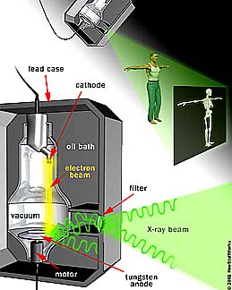
site. X-rays are produced when
electrons are impelled against an anode metal target (tungsten; copper; molybdenum;
platinum; others) as they pass through a vacuum tube at high speeds driven by
voltages from 10 to 1000 kilovolts (kV). When incoming electrons interact with
inner electrons in the metal, these latter are driven momentarily to higher
energy levels (these orbital electrons are pushed into outer orbitals); when
these excited electrons drop back to their initial orbits (a transition from
a higher to a lower energy level), the energy they acquired is given off as
radiation, including x-rays . Some of the scattered x-rays are collimated into
beams (typically at conical angles up to 35°) that are directed towards targets
(such as the human body). Soft body tissue absorbs less x-rays, i.e., passes
more of the radiation, whereas bone and other solids prevent most of the x-rays
from transmitting through the body mass. (X-rays have other uses, such as examining
metals for flaws or determining crystal structure.) Here is a diagram of a typical
x-ray machine setup:
Two classes of detectors
record the x-ray-generated image: 1)Photographic film, in which the difference
in gray levels or tones relates to varying absorption of the radiation in the
beam impinging on the target (the convention is to use the exposed film [x-rays
act on the silver halide {see page 12 of this Introduction} to reduce it to
metal silver grains] in its negative form, such that bone will appear nearly
white [thus, because bone absorbs efficiently, few x-rays strike the corresponding
part of the film, leaving it largely unexposed; the soft tissue equivalents
pass much more radiation and darken the film]); 2) fluorescent screens, that
include phospors (elements or compounds that fluoresce or phosphoresce) coating
a substrate; this occurs when electrons in the phosphors jump to higher level
orbitals, with visible light given off either instantly when the electrons transition
back to the lower state or with a time delay fractions of a second or seconds
(afterglow), in a process similar to x-ray production; typical phosphors include
Calcium tungstate or Barium Lead sulphate (many other compounds are available
such as Lead oxide or those containing Gadolinium or Lanthanum; these screens
in certain configurations allow realtime movements of the medical patient to
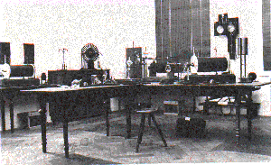
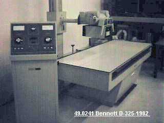
be observed and the sreen images can be photographed or digitized. Here is a typical hospital
examining room that contains the setup used in x-ray radiology; the table on
which the patient lies that can, in some instruments, be raised to a vertical
position.
X-ray radiology is still
the most commonly used medical instrument technique. Here are a sequence of
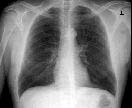
images that illustrate typical uses and results. The first is a chest x-ray,
(the skeletal bones are whitish since they absorb the radiation and thus the
negative is not darkened and the lungs dark because more of the radiation has
passed through them):
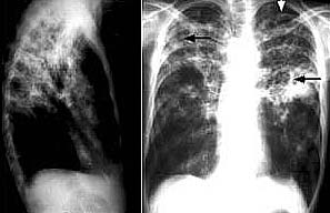
This next is a front and
side view of the upper torso; the arrow points to a tuberculosis patch in the
left lung:
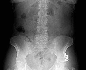
Here is a negative x-ray
film image of the pelvic area:
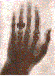
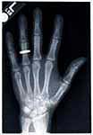
Compare this recent image
of the human hand with that shown above as the first ever taken:
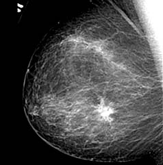
This next picture is a
mammogram showing a growth in the female breast:
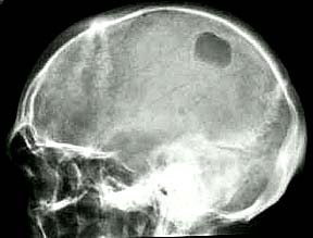
The human skull is x-rayed
mainly to spot signs of fracture. But, sometimes indications of tumors are present,
as shown by the darker gray patch in the cranium of this individual's skull:
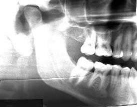
The jaw and teeth are evident
in this lateral view of the lower human skull:
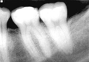
Most of us gain our first
experience and insight into x-rays when we have a small film inserted into our
mouth and then the x-ray machine is placed against that part of our jaw. Here
is a typical x-ray image of teeth, in which the whitest part of the negative
corresponds to metal fillings:
An important variation
in x-ray radiography is Fluoroscopy. In this method, either chemicals
that react with x-rays are swallowed or inserted as an enema or chemicals/dyes
are injected into the blood stream. These tend to increase the contrast between
soft tissue response in the parts of the body receiving these fluids and surrounding
bone and tissue. This pictorially highlights abnormalities. Barium sulphate is a good
example. When swallowed (either at once or commonly in gulps), the "Barium Cocktail"
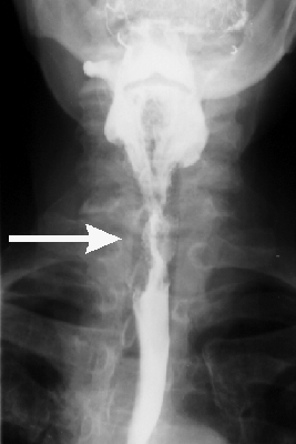
is especially useful in examining the digestive track. In this image, an obstruction
in the esophagus carrying food and liquids into the stomach is made evident:
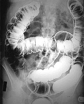
The large intestine or
colon is strikingly emphasized in a patient who has just received a Barium enema:
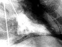
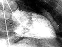
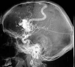
Still another variant is
the Angiogram. This involves insertion of a catheter into an artery,
accompanied by a dye that reacts to x-rays. It is commonly used to explore the
areas in and around the heart. Here is a pair of views of the left ventricle
of the heart when it is pumping and squeezing blood and thus contracting (systolic
phase) and then expanding as blood is returned (diastolic phase):
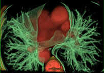
This next image is an angiogram
that has been colored to show blood vessels including the great trunk artery
or aorta around the heart: Using special methods,
angiogram-like images can be made for the blood vessels in the human head:
We move on now to a powerful
new approach to medical imaging, based on the technique of tomography,
which uses computers to assist in obtaining three-dimensional images or image
slices when either x-rays or radioactive elements (nuclear medicine) are involved
in producing radiation-based imagery.