

| Introduction to
the Malformed Amphibian Issue Return to NARCAM Contents |
In the summer of 1995, middle school students on a field trip to a farm pond in southern Minnesota discovered large numbers of frogs with misshapen, extra, or missing limbs. About 50% of the northern leopard frogs they caught that day were malformed. Since then, there has been a dramatic increase in reports of malformed amphibian in North America. To date NARCAM has received over 2100 reports from 1032 sites encompassing 82 species of amphibians. Of these, there are 944 reports with verifiable cases of malformations involving 52 species in 46 states and 4 provinces.
However, malformations seem to be concentrated in a few core species, with the Northern Leopard Frog (Rana pipiens) being most commonly reported (30.1% of all reports of malformations). Reports of malformed Green Frogs (R. clamitans), American Bullfrogs (R. catesbeiana) and Pacific Treefrogs (Pseudacris regilla) are fairly frequent as well (13.1%, 10.0% and 9.3% respectively of all reports), and these four species plus an additional six (Bufo americanus, R. sylvatica, Hyla chrysoscelis & versicolor, R. septentrionalis and B. boreas) account for 77.8% of all reports. Also, while 78.8% of all reported species showing malformations were anuran amphibians, anurans accounted for 94.1% of these reports. (See Results page for a more detailed analysis of the data.)
Although historical records on amphibian populations are limited, what records that do exist suggest that observations of malformations in amphibians is not a new phenomenon. Nevertheless, observations from researchers and collectors that have been working with amphibians for many years and a growing number of multi-year studies indicate that malformations may be an unusual and recent phenomenon in parts of the U.S.A. and Canada (Reaser 2000). While some controversy exists over the extent to which these malformations occur naturally in healthy populations, with most estimates at 2% or less (Ouellet et al. 1997, Gardiner & Hoppe 1999, Sessions et al. 1999), scientists agree that current numbers of reported malformations are in excess of what is likely to be normal and that the situation warrants urgent attention (Meteyer et al. 2000). The wide geographic distribution and the variety of amphibian malformations are a growing concern to resource managers, research scientists and public health officials.
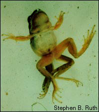 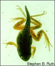  |
Examples of malformed Pacific Treefrogs (Pseudacris
regilla) |
The potential for malformations to serve as a sign of ecosystem disruption, and the effect this potential disruption might have on other organisms that share those ecosystems has not yet been resolved. Malformations represent an error that occurred early in the process of development and investigations into the causes of these malformations have focused on agents that could disrupt this process (Meteyer 2000). Field and laboratory evidence suggests that three major types of environmental agents can induce malformations in developing frogs: UV-B radiation, chemicals, and parasites.
UV-B Radiation. There has been much concern about atmospheric ozone depletion and increased levels of UV-B radiation reaching the ground. Laboratory experiments have shown that continuous exposure of developing frog embryos to UV-B results primarily in bilaterally reduced limbs, i.e. reduced or missing limb segments that appear as blunt-ended truncations (Ankley et al. 1998).
There is no evidence, however, that UV-B is a direct cause of amphibian limb malformations in the field. The truncated limbs found in wild frogs are usually only on one side and tapering, rather than bilateral and blunt-ended (Gardiner & Hoppe 1999). Furthermore, most amphibian eggs are protected from UV-B by their natural pigment and shade provided by vegetation in and around the pond. There is as yet no evidence that these types of malformations are more prevalent in more open ponds, or ponds at high altitudes where exposure to UV-B would be greater. Proof that UV-B can be a direct inducer of limb abnormalities in the field will require very careful replication in the laboratory of intensities of UV-B radiation typical of field conditions. UV-B radiation in the field might act in a more subtle manner, however, by activating photosensitive compounds or breaking them down into products that cause malformations, as discussed below.
Chemical Agents. The use of pesticides has been correlated with anuran hind limb deformities in the St. Lawrence river valley in Quebec (Ouellet et al. 1997). Newly metamorphosed frogs in ponds near farmland treated with pesticides have a high incidence of reduced limbs compared to frogs in ponds surrounded by untreated land. The reduced limb malformation was reproducible in the laboratory by continuous exposure of Xenopus embryos to water, sediments, and/or aqueous sediment extracts from ponds with high incidences of malformations (a so called Frog Embryo Teratogenesis Assay-Xenopus, or FETAX; Fort et al. 1999a). The effects were also dose-dependent.
Certain limb malformations can be induced in the laboratory by retinoids, giving rise to speculation that they could be the agents causing this malformation in wild frogs. Retinoic acid, a lipid-soluble derivative of vitamin A, is a hormone that controls limb development in all vertebrates and limb regeneration in amphibians. Retinoids can induce multiple limbs or limb segments in regenerating amphibian limbs (Niazi 1996, Maden 1998). Retinoids also induce the type of bone bridges observed in the malformed hind limbs of wild frogs (Gardiner & Hoppe 1999, Meteyer et al. 2000).
In contrast, retinoids inhibit anuran hind limb bud development, causing reduced limbs (Niazi 1996). The effects of retinoids on hind limb development has a temporal component. Early exposure yields limbs with a short or missing femur or lower leg (tibiafibula), but with normal foot. Later exposure results in limbs missing the foot or with missing or reduced digits (ectrodactyly or brachydactyly).
So far, however, it has not been shown that retinoids are the cause of malformations in the wild. If that was the case, they should be detectable in the water and/or sediments of ponds, or in the algae eaten by tadpoles. Most retinoids are insoluble in water, making it difficult to apply them to tadpoles and embryos in the lab, so it is difficult to envision how they could affect tadpoles directly through the water in a pond, unless there are other substances present that make them soluble.
Methoprene, a photosensitive insecticide, is broken down by UV-B radiation into compounds that mimic the structure and binding properties of retinoic acid (La Clair et al. 1998). These breakdown products have induced neural, craniofacial, axial, and gut defects in a FETAX assay, implying that they are at least partially soluble in water. One of the break-down products was identified by mass spectrometry in the water of three sites in Vermont with high levels of malformed frogs, but at levels much lower than was required to cause deformities in the FETAX assay (La Clair et al. 1998). Methoprene had been applied several months earlier, so it is possible that tadpoles were exposed to higher levels of the breakdown product prior to the study.
Carbaryl, another insecticide, greatly increased mortality of frog embryos and tadpoles in the laboratory when exposed to UV-B light at levels below those normally reaching the ground (Zaga et al. 1998). Xenopus laevis embryos and tadpoles are also very sensitive to increased concentrations of the bivalent ions of zinc, copper, nickel, cobalt, and cadmium. Abnormally high concentrations of these ions cause a variety of malformations, including reduced limbs (Luo et al. 1993, Plowman et al. 1994).
Another aspect to consider is that chemical agents in solution are likely to affect the entire individual, including the limbs on both sides. Under such circumstances one would expect that the malformations induced would be symmetrical, which has been shown in experimental studies (Sessions et al. 2001). However, some scientists dispute that this would exclusively be the case and believe that chemical agents could under the right conditiond induce asymmetrical malformations. The issue is far from academic - field-caught samples of malformed amphibians most often show asymmetrical malformations. The possibility that chemical agents could induce asymmetrical malformations in the field has not been ruled out and at this point it would be premature to assume that chemical agents could only induce symmetrical malformations.
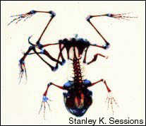
Parasites. Multiple limbs are the predominant malformation exhibited by wild frogs at some sites. This phenotype is correlated with infection of the frogs by larvae of the trematode flatworm Ribeiroia (Sessions & Ruth 1990, Johnson et al. 1999, Sessions et al. 1999). The collection sites in the study of Johnson et al. (1999) tested negative for pesticides, PCBs, and heavy metals, and frog eggs collected from these sites developed normally under laboratory conditions. Ribeiroia eggs are released from their hosts (presumably birds) and picked up by snails. After growth in the snails they develop into swimming larvae that find their way preferentially into the developing pelvic girdles and hind limb buds of frog tadpoles, where they form cysts.
Johnson et al. (1999) showed that Ribeiroia cysts can cause limb malformations by infecting frog tadpoles in the laboratory with various concentrations of trematode larvae (cercariae) similar to the densities found in infected wild frogs. Hind limbs malformations developed in 85% of the total number of experimental frogs that survived to metamorphosis. Survival decreased to 40% at the highest concentration of trematode larvae, and all of the surviving frogs at that concentration exhibited hind limb malformations. Exposure of frogs to Alaria larvae, another species of trematode found in frog breeding ponds, caused no increase in mortality or limb abnormalities. However, multilegged frogs with no obvious evidence of trematode infection have been found in many locations, so it is not likely that trematode cysts are the explanation for all multiple limbed frogs.
Tetrapod limb development is controlled by three interdependent signaling centers in the limb bud, and each of these centers produces specific sets of signaling molecules that together set up the three-dimensional pattern of the limb (Vogt & Douboule 1999). Thus limb development is altered by environmental agents when they interfere with the production or activity of one or more of these signaling molecules and the feedback loops of which they are part.
Retinoic acid is one such signaling molecule, and excess amounts of this chemical or one of its mimics can cause a failure of one or more signaling centers and thus reduced limbs, the nature of the reduction being dependent on the stage of development at which exposure occurred.
Trematode cysts could cause the formation of multiple limbs via mechanical means, creating multiple sets of signaling centers. But why do Ribeiroia cysts cause multiple limbs and not Alaria cysts? Most likely because Alaria larvae show no site preference when infecting tadpoles and thus their cysts are widely distributed throughout the subcutaneous tissue of the frogs, causing minimal mechanical damage, whereas Ribeiroia larvae concentrate in the pelvic and hind limb regions where the hind limb buds receive the brunt of the damage (Johnson et al. 1999). Missing limbs might also be explained by the complete destruction of limb tissue by trematode infection.
What is the sum of our knowledge of the causes of amphibian malformations in the wild? The water and sediments of ponds where the reduced limb malformations predominates clearly contain agents that induce this phenotype in laboratory assays. Reduced limbs can be induced in the laboratory by retinoids and there is evidence that retinoids or retinoid-like compounds may be responsible for what is seen in the field. Trace amounts of one methoprene breakdown product have been reported from three frog breeding sites, but there is as yet no proof that retinoids or retinoid-like compounds are present in ponds at concentrations that can induce developmental abnormalities.
Also, UV-induced retinoid-like breakdown products of the insecticide methoprene have been shown to cause craniofacial and axial malformations in Xenopus embryos. Trematode infections can cause multiple limb malformations in the laboratory and are strongly associated with this malformation in the nature. Yet this malformation can also be found in locations with no obvious evidence of trematodes.
It is unlikely that we have identified all the potential agents that can cause developmental abnormalities in frogs. A large number of industrial chemicals and chemicals present in animal and plant waste are finding their way into the environment, many of which might be candidates for causing amphibian developmental malformations. In addition, we still know little about the multitude of natural agents that might affect amphibian development, such as nutrition, pH, chemicals produced by plants and/or other animals, microbes, and overcrowding, for example. Furthermore, natural or anthropogenic environmental factors that are not by themselves toxic could interact with one another in synergistic ways to cause developmental abnormalities. Compounds that are not toxic in one form could become toxic through the action of sunlight, water and metabolic passage through a variety of organisms (Stocum 2000).
Clearly, the available data is insufficient to draw firm conclusions about the causes of malformations in wild frogs. The data does suggest that the wide variety of malformations encountered at different sites cannot be attributed to a single common cause (Meteyer et al. 2000). We also know little about how anthropogenic and natural environmental agents affect amphibian development in the wild. Thus each site where malformations are found must be carefully studied, and we need to learn much more about how these agents affect, and are affected by the ecology of the regions where frogs live. There is also increasing recognition of the need for multidisciplinary approaches to illuminate how environmental changes affect the ecological parameters (and vice versa) within which anuran development takes place (Stocum 2000).
Over the last 100 years or so, many species of amphibians (frogs, toads, salamanders and newts) throughout the world have declined markedly in numbers. Some species have become extinct. In many cases the declines are a direct response to the impact of human activities that result in habitat destruction. In addition, towards the late 1980's, biologists began realizing that amphibian populations in apparently pristine habitats in many different parts of the world were experiencing population declines. This led to the suggestion that there may be one or more global factors that are adversely affecting amphibians. A more thorough discussion of this issue can be found on the Declining Amphibian Populations Task Force (DAPTF)'s web site.
Without a clear understanding of causes of amphibian malformations, the link between malformations and widespread amphibian declines remains uncertain. Nevertheless, the fact that most malformations have been found in recently metamorphosed individuals suggests that malformed amphibians often do not survive into adulthood. Thus at a local scale malformations may contribute to the decline of populations, but is unlikely to explain the disappearance of species worldwide (Reaser 2000). For example, no malformation crisis has been reported in Europe to date, yet many species there are in a severe decline. Still, amphibian malformations should be a part of the amphibian decline discussion for the following reasons:
In cooperation with the scientific community, this web site has been designed as an avenue for people to report sightings of amphibian malformations in North America. Regardless of whether you have observed malformed amphibians, or you have handled a number of wild amphibians but without noting any malformations, we urge you to report your sightings by using one of our online submission forms (there is a technical form for biologists and a form for non-biologists). If you would like information on the extent of malformation reports we have received, go to Where Have Malformations Been Reported? There you will find the geographic distribution of reports as well as information on the types of malformations found and the species affected in each area.
The data gathered at this web site will help inform the public, offer a resource to scientists investigating malformations, and provide key information to land managers. The effectiveness of compiling these data depends on your accurate reports -- reports of both malformed and normal amphibians. Knowing where malformations are not a problem is just as important as knowing where they are. Your reports can speed up the process of linking malformations to a cause or group of causes. Please enter your name, address, and phone number at the top of the Survey Form, as indicated, so we can contact you about your report. If appropriate, we will inform a herpetologist or wildlife biologist in your area who could help with a follow-up survey and collect additional data at the site you visited.
PAPERWORK REDUCTION ACT statement: The information collected will be used by scientists and agencies to identify areas where malformed amphibians occur and the rates of occurrence. Correlating that information with other information about hypothesized causes of malformations may lead to better analyses of those features. Participation in this data collection effort is voluntary. An agency may not conduct or sponsor a collection of information unless it displays a currently valid OMB control number. The currently valid OMB control number of this survey is 1028-0056.
Burden Estimate Statement: Public burden for reporting information requested for this form is estimated to average 20 minutes per respondent. Comments regarding this collection of information may be directed by mail to the USGS Information Clearance Office, 12201 Sunrise Valley Drive MS 807, Reston VA 21092.
Ankley G.T., Tietge J.E., DeFoe D.L., Hjensen K.M., Holcombe G.W., Durhan E.J. & Diamond A. 1998. Effects of ultraviolet light and methoprene on survival and development of Rana pipiens. Environ Toxicol Chem 17: 2530-2542.
Fort D.J., Rogers R.L., Copely H.F., Bruning L.A., Stover E.L., Helgen J.C. & Burkhart J.B. 1999. Effects of pond water, sediment, and sediment extracts from Minnesota and Vermont, USA, on early development and metamorphosis of Xenopus. Environ Toxicol Chem 18: 2305- 2315.
Gardiner D.M & Hoppe D.M. 1999. Environmentally induced limb malformations in mink frogs (Rana septentrionalis). J Exp Zool 284: 207-216.
Johnson P.T.J., Lunde K.B., Ritchie E.G. & Launer A.E. 1999. The effect of trematode infection on amphibian limb development and survivorship. Science 284: 802-804.
La Clair J.J., Bantle J.A. & Dumont J. 1998. Protoproducts and metabolites of a common insect growth regulator produce developmental abnormalities in Xenopus. Environ Sci Technol 32:1453-1461.
Luo S.Q., Plowman M.C., Hopfer S.M. & Sunderman F.W. 1993. Embryotoxicity and teratogenicity of CU21 and Zn21 for Xenopus laevis, assayed by the FETAX procedure. Ann Clin Lab Sci 23: 111-120.
Maden M. 1998. Retinoids as endogenous components of the regenerating limb and tail. Wound Rep and Reg 6: 358-365.
Meteyer C.U. 2000. Field guide to malformations of frogs and toads with radiographic interpretations. Biological Science Report USGS/BRD/BSR-2000-0005. (See: What Do the Malformations Look Like?)
Meteyer C.U., Loeffler I.K. , Fallon J.F., Converse K.A., Green E., Helgen J.C., Kersten S., Levey R., Eaton-Poole L. and Burkhart J.G. 2000. Hind limb malformations in free-living Northern Leopard Frogs (Rana pipiens) from Maine, Minnesota, and Vermont suggest multiple etiologies. Teratology 62: 151-171.
Niazi I.A. 1996. Background to work on retinoids and amphibian limb regeneration: studies on anuran tadpoles-a retrospect. J Biosci 21: 273-297.
Ouellet M., Bonin J., Rodrigue J., DesGranges J-L. & Lair S. 1997. Hind-limb deformities (ectromelia, ectrodactyly) in free-living anurans from agricultural habitats. J Wildl Dis 33: 95-104.
Plowman M.C., Grbac-Ivankovic S., Martin J., Hopfer S.M. & Sunderman F.W. 1994. Malformations persist after metamorphosis of Xenopus laevis tadpoles exposed to Ni21, Co21,or Cd21 in FETAX assays. Teratog Carcinogen Mutagen 14: 135-144.
Reaser J.K. 2000. Amphibian declines: an issue overview. Federal Taskforce on Amphibian Declines and Deformaties, Washington, DC.
Sessions S.K., Franssen R.A. & Horner V.L. 1999. Morphological clues from multilegged frogs: are retinoids to blame? Science 284: 800-802.
Sessions S.K. & Ruth S.B. 1990. Explanation of naturally occurring supernumerary limbs in amphibians. J Exp Zool 254: 38-47.
Sessions S.K., Stopper G., Hecker L., Horner V. & Franssen A. 2001. Deformed Amphibian Research at Hartwick College. www.hartwick.edu/biology/def_frogs/ (meth/prob1.html).
Stocum D.L. 2000. Frog limb deformities: an "eco-devo" riddle wrapped in multiple hypotheses surrounded by insufficient data. Teratology 62: 147-150.
Vogt T.F. 1999. Antagonists go out on a limb. Cell 99: 563-566.
Zaga A., Little E.E., Rabeni C.F. & Ellersieck M.R. 1998. Photoenhanced toxicity of a carbamate insecticide to early life stage anuran amphibians. Environ Toxicol Chem 17: 2543-2553.
